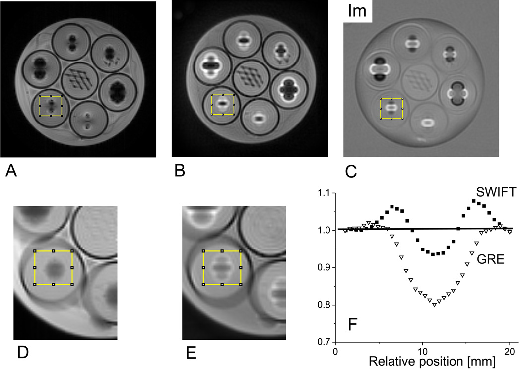
Figure 1.
Imaging studies of a phantom consisting of a plastic jar filled with saline and 7 tubes containing gelatin, titanium balls, and plastic mesh. The images are magnitude mode GRE (A), magnitude mode SWIFT (B), and imaginary mode SWIFT (C). The slice shown contains the titanium balls in the outer 6 tubes and the plastic mesh in the center tube. The balls are situated at slightly different heights; therefore, the slice shown does not cut through the center of every ball. The diameters of the titanium balls in the clockwise direction starting from the top are: 3.97, 2.38, 3.18, 4.76, 2.38 and 3.18 mm. The rims of the tubes are clearly identifiable from the black edge. The displaced and piled-up signals around the balls are clearly visible in the SWIFT images. Expanded views of the GRE and SWIFT images obtained by averaging 20 adjacent slices are also shown with the volume-of-interest (yellow box) from which the summed profiles (F) were obtained.

