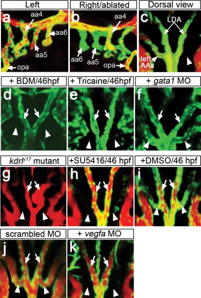
Figure 1. AA5x angiogenesis requires flow and Vegf signaling.
a-k, Tg(kdrl:egfp)la116 embryos at 72 hpf (a-c), 60 hpf (d-f) or 65 hpf (g-k). a-c, g-k Embryos subjected to microangiography. Endothelial cells are green, flow is red. a, b, Lateral views, anterior to left (a), or right (b), dorsal is up. c-k, dorsal view, anterior is up. a, b, aortic arches (AA, numbered, indicated by arrows) after severing right AA5 and 6 from ventral aorta; opa – opercular artery. c, dorsal view of embryo in a, b. d, e, Stills from Supplementary Movies 5 and 6. Embryos treated beginning at 46 hpf with BDM (d), Tricaine (e), or injected with gata1 MO (f). g, kdrly17 mutant embryo at 65 hpf. h-k, Embryos treated with 2.5 μM SU5416 (h) or 0.1% DMSO (i) beginning at 46 hpf or injected with 3 ng of scrambled MO (j) or 3 ng of Vegfa MO (k). c-k, Arrows: lateral dorsal aortae (LDA), arrowheads: AA5x.

