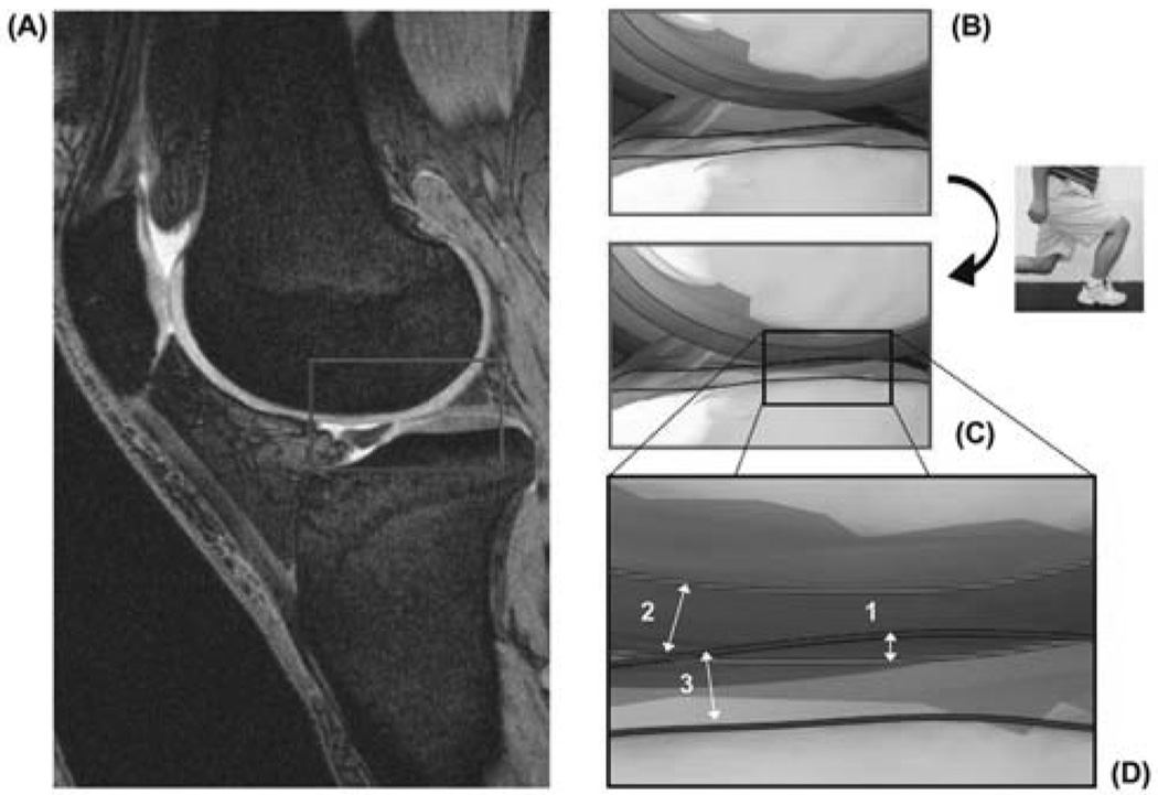
Figure 1.
Illustration of the calculation of compartmental contact deformation. The MR images of the knee joint after ~1 hour non-weight bearing (A) are used to determine the respective cartilage thicknesses of the femur (top) and tibia (bottom) at rest (B). After matching the MR models to the fluoroscopic images captured during weight bearing lunge (C), compartmental cartilage deformation is calculated by dividing the amount of penetration (1) by the sum of the femoral (2) and tibial (3) cartilage surface thicknesses, as illustrated in (D). (Reproduced from: Bingham JT et al. Rheumatology (Oxford). 2008 Nov;47(11):1622-7. Reprinted with permission)

