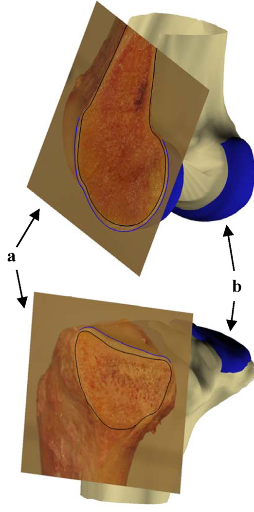
Figure Appendix.
Illustration of the comparison of direct measurement of cartilage thickness on cadaveric cross-sections with that on MR image based models. The digital images of the cadaver osteotomies (a) were matched to the respective cross-section planes of the MR image models (b). The black (bone mesh) and blue (cartilage mesh) lines on the digital images indicate the intersection of the cartilage mesh models with the digital image at the location of osteotomy. Only two cross-sections are shown for illustrative purposes.

