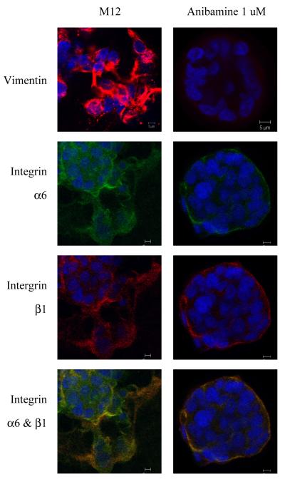
Figure 7.
Comparison of content and localization of vimentin and integrin within the morphological structures formed by the M12 prostate sublines +/− anibamine grown embedded in lrECM gels. Confocal immunofluorescence microscopy of structures formed at day 8 stained with antibodies to vimentin (red, top panel), α6-integrin (green) and β1-integrin (red) as indicated. The overlay of α6β1-integrin is shown on the bottom panel. All pictures are taken at a magnification of 63X and nuclei were counterstained with 4′6-diamidino-2-phenylindole (DAPI; blue) as discussed in Materials and Methods.

