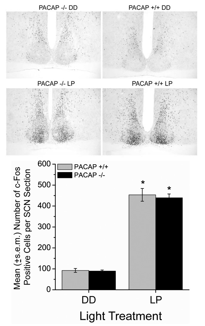
Figure 5.
Loss of PACAP does not alter light induction of c-Fos protein in late night. Representative photomicrographs (10× magnification) of the medial SCN show similar patterns and numbers of c-Fos positive cells in PACAP +/+ and −/− mice at CT 22 under basal (top), and light-stimulated (10-min, ~100-lux light pulse, middle) conditions. Graph shows mean (+/− s.e.m.) number of c-Fos immunoreactive cells per unilateral SCN section (40 µm, medial SCN). * p < 0.0001, vs DD, Two-Way ANOVA, Tukey post-hoc.

