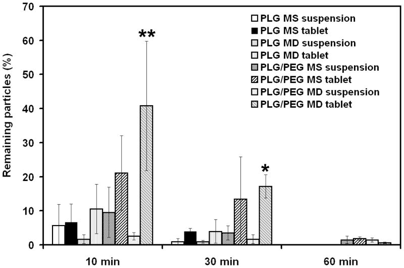
Figure 6.
In vivo residence time of microparticles on the rabbit eye. The percentage of microparticles remaining on the preocular surface of rabbits was measured for eight different microparticle formulations. **At 10 min, PLG/PEG MD tablet was significantly different from all other formulations (p < 0.05) except PLG/PEG MS tablet. *At 30 min, PLG/PEG MD tablet was significantly different from all suspensions (p < 0.05). The data points represent the average of n = 3 – 5 measurements ± standard deviation.

