
Fig. 2.
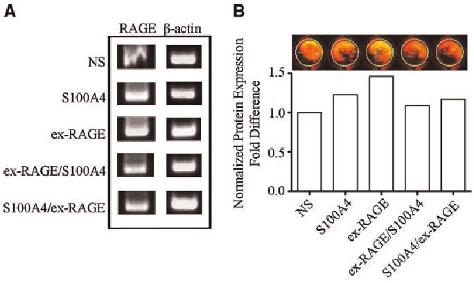
Inhibition of S100A4-induced RAGE gene expression by ex-RAGE. A: The greatest gene expression was demonstrated by RT-PCR for HSG cells treated with ex-RAGE alone (50 ng/ml). A decreased gene expression was observed when ex-RAGE was added prior to or after S100A4 treatments (separated by a 30 min interval). B: Protein expression of RAGE was examined by In-Cell Western analysis using the same experimental design for RT-PCR analysis. Following a rabbit anti-human RAGE primary antibody incubation (1: 100 dilution), the cells were treated with an anti-rabbit IgG secondary antibody (1: 1,000 dilution) conjugated with IRDye800CW. The absorbance values were normalized for analysis.