
Fig. 3.
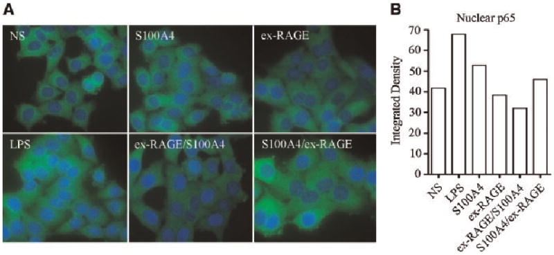
Inhibition of NF-κB p65 translocation by ex-RAGE. A: The activation status of NF-κB p65 subunit was examined by immunocytochemistry. HSG cells were stained for p65 (green) following treatments and counterstained with Hoeschst Dye (blue). B: Quantitative analysis using ImageJ, which measures green fluorescence intensity in the nucleus, indicated an inhibitory response of NF-κB p65 translocation in ex-RAGE/SI00A4 treated HSG cells.