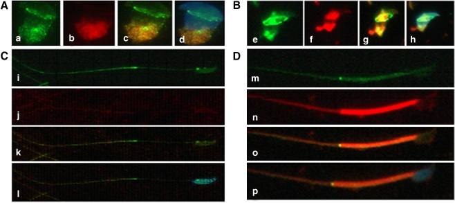
Fig. 2.
Immunofluorescence assay of SEPT7 (green) and mitochondria (MitoTracker, red) in developing spermatozoa (I–IV). I and II (a–h) indicate early spermiogenesis. In the round spermatid (I, a–d), mitochondria are localized at the opposite side of the nucleus. With advancing spermiogenesis (II, e–h), the tail starts to develop and the mitochondria are divided into two parts. With the elongation of the tail (III, i–l), the mitochondria are diffused into the tail region. At this stage, SEPT7 was localized predominantly at the neck and the annulus. In mature sperm (IV, m–p), SEPT7 appeared mainly as a bright dot signal at the annulus. It also exhibited a punctate pattern at the mitochondrial sheath and at the head. The nucleus was stained with DAPI (blue)

