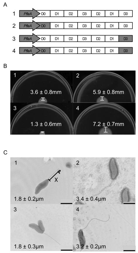
Figure 2. Motility assays and TEM of domain swaps.
Panel A shows a linear schematic of the domain organization of the C. jejuni flaB gene and three constructs containing the domain swaps (not drawn to scale). Regions in gray correspond to C. jejuni flaA gene, while regions in white correspond to the C. jejuni flaB gene. Motility assays and TEM of flagellar mutants transformed with a vector harboring flaB, which had been modified to include the D0 domain of FlaA. C. jejuni strains were cultured on MH 0.4% agar plates, and the distance from the edge of the culture spot to the haze of motility was determined (Panel B). Bacteria were stained with 1% phosphotungstic acid, TEM performed, and the length of the flagellum was measured (Panel C). The images are of the: 1) C. jejuni F38011 flaAB mutant harboring PflaA-flaB; 2) C. jejuni F38011 flaAB mutant harboring PflaA-flaAD0BD123BD0; 3) C. jejuni F38011 flaAB mutant harboring PflaA-flaBD0BD123AD0; and 4) C. jejuni F38011 flaAB mutant harboring PflaA-flaAD0BD123AD0. The values shown represent the mean ± standard deviation of 6 motility assays or 10 TEM images. One representative image is shown for each strain. X = Example of line drawn to measure flagellar length. Bar = 1 μm.

