
Figure 2.
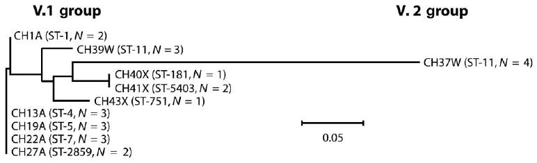
Phylogram of the factor H–binding protein (fHbp) polypeptide sequences from African strains. The relative distance between respective peptides is shown on the horizontal line; the scale bar denotes 5 changes/100 amino acids. One fHbp amino acid sequence from each sequence type (ST) is represented, except for ST-11 (group W-135), for which data from strains with fHbp variant 1 (v.1) or 2 (v.2) are shown. N, the no. of isolates with identical capsular group, ST, and fHbp polypeptide sequence. Peptide identifiers for each strain are shown in table 1.