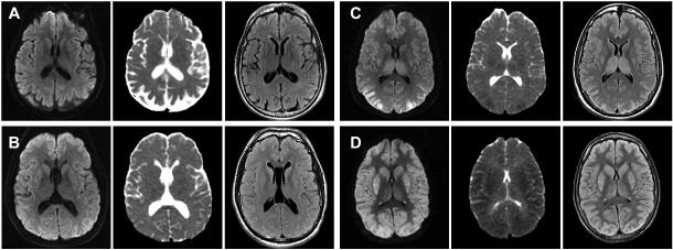
Figure 2.
Brain MRI (DWI, ADC, and FLAIR) of two patients in the good (A, B) and two patients in the poor (C, D) outcome groups between 55 and 99 hours after the arrest. The cortical and deep gray structures were qualitatively rated as normal and possibly abnormal in A, possibly and mildly abnormal in B, moderately and severely abnormal in C, and all severely abnormal in D.

