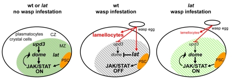
Figure 7. Model for lat function in Drosophila larval hematopoiesis.
During normal development (left panel), PSC cells (orange) act, in a non–cell-autonomous manner (arrow) to maintain JAK/STAT signalling activity and preserve a pool of multipotent prohemocytes in the MZ (green shades). The PSC signal overrides lat function in the MZ (grey shades). In response to parasitisation (middle panel), there is a decrease of upd3 and dome and increase of lat transcripts, which ultimately lead to an increased lat/dome ratio. The PSC signal is short-circuited. As a result, JAK/STAT signalling is switched off, thus licensing prohemocytes to differentiate into lamellocytes. Lat activity is strictly required in the LG for this switch. In the absence of lat (right panel), residual upd3 levels maintain JAK/STAT activity, therefore preserving a pool of prohemocytes (grey shades). Upon wasp parasitisation some differentiating hemocytes become lamellocytes, however, indicating that lat is not required for this differentiation program per se. Arrows indicate activation, vertical bars repression.

