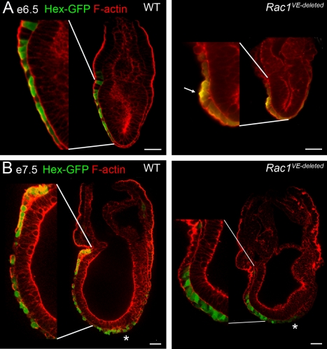
Figure 4. Epithelial organization of Rac1 VE-deleted embryos.
Individual confocal sections of whole-mount embryos stained for F-actin (red). AVE cells were marked with the Hex-GFP transgene (green, staining with anti-GFP antibody in A and native GFP in B). After AVE migration is complete, at e6.5 (A) and e7.5 (B), AVE cells formed a single-layer epithelium and were squamous in wild-type embryos, with the exception of the most proximal cells, which were cuboidal. At e7.5, definitive endoderm cells expressing Hex-GFP (marked by an *) were present at the distal end of the primitive streak. In VE-deleted embryos, AVE cells had failed to reach the embryonic/extra-embryonic boundary. The Hex-GFP-expressing cells retained the columnar shape characteristic of migrating AVE cells and displayed a strong apical actin at e6.5 (arrow in A). Scale bars = 50 µm. Insets are 2×.

