Fig. 4.
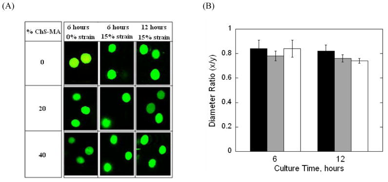
(A) Representative confocal microscopy images of live chondrocytes (green) encapsulated in PEG-only or PEG-ChS gels and subjected to either not strain or a gross unconfined, 15% static compressive strains. Gels were imaged before (at 6 hours) being placed into the bioreactor and after (at the first 12 hours of culture) being subjected to dynamic loading. Prior to placing in the bioreactor, cellular morphology was generally round, but with the application of a 15% strain, the cells adopted an oblate ellipsoid morphology for all gel formulations before and after being subject to loading; Original magnification is 40×. (B) Chondrocyte deformation was quantitatively assessed under a gross, unconfined, 15% static compressive strain before and after dynamic loading for PEG-only (black bars) and PEG-ChS gels containing 20% ChS-MA (gray bars) and 40% ChS-MA (white bars). Cellular deformation was quantified by a diameter ratio (x/y) defined by the ratio of the cell diameter parallel to the direction of strain (x) and the cell diameter perpendicular to the direction of the applied strain (y) measured at full width half maximum height; **p<0.01.

