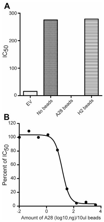
Fig. 5.
Adsorption of VACV neutralizing antibody. (A) Samples of pooled serum following the fourth A28/H2 DNA immunization were incubated with A28 or H2 coated magnetic beads and the depleted sera tested for ability to neutralize VACV. Averages of duplicate determinations are shown. EV, refers to empty vector. (B) Magnetic beads were coated with five-fold serial dilutions of A28 protein, incubated with pooled A28/H2 sera, and the depleted sera tested for VACV neutralization as in panel A. The percentage of neutralizing activity remaining after adsorption is plotted. Averages of duplicate determinations are shown.

