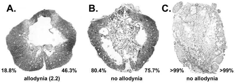
Figure 7.
Photomicrographs taken at the epicenter of spinal cord contusion injuries from allodynic and non-allodynic animals. The percentage values indicate the amount of area 3 damage (re Figure 1) on each side of the spinal cord. Magnification factor of 40X.

