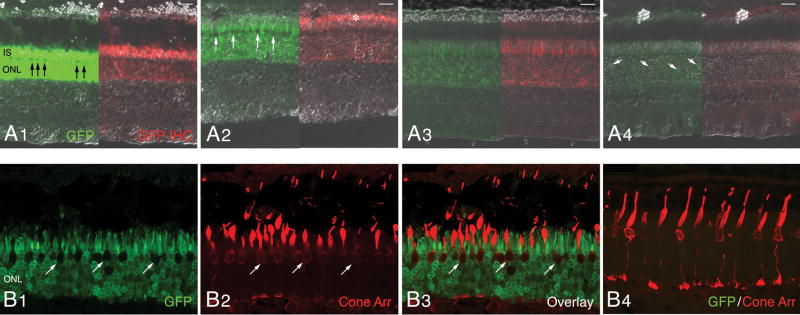
Figure 4. Transduction pattern of AAV2/5-mOP-GFP following subretinal delivery to the normal canine retina.
Native GFP fluorescence (green) and GFP immunolabeling (red) images merged with DIC/Nomarski optics; images show the GFP expression with decreasing viral vector titers (A1–4). A1) Retina of E1048-R (3.27 × 10¹³ vg/ml). A2) Retina of E1048-L (3.27 × 10¹² vg/ml). A3) Retina of P1473-R (3.27 × 10¹¹ vg/ml). A4) Retina of P1473-L (3.27 × 10¹⁰ vg/ml). Confocal microscopy images of native GFP (green) and cone arrestin (red) immunolabeling in the injected area (B1–3) and non-injected area (B4) of retina E1048-R.
Abbreviations: IS, inner segments; ONL, outer nuclear layer; GFP, green fluorescent protein; IHC, immunohistochemistry; Cone Arr. cone arrestin.
Scale bars: (A1–4) 20 μm; (B1–4) 25 μm.

