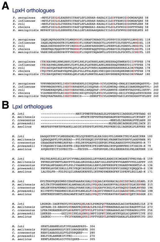
Figure 2.
Sequence alignments of LpxH and LpxI orthologues. Panel A. Sequence alignments of five representative LpxH orthologues. Panel B. Alignments of five typical LpxI orthologues. In each panel, the absolutely conserved residues are colored red. There is no conservation of domains or of possible active site motifs between LpxI and LpxH, which are members of distinctly different protein families. The sequences were obtained from (http://www.ncbi.nlm.nih.gov/sutils/genom_tree.cgi).

