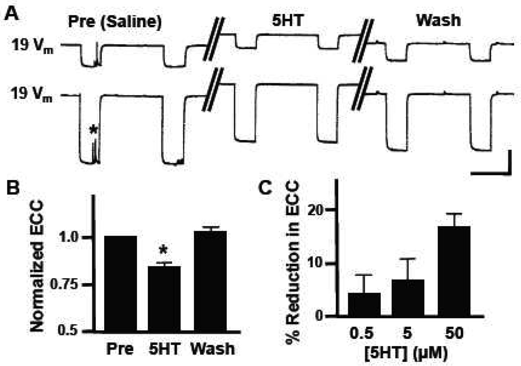
Figure 1.
Neuromodulatory effects of serotonin on 19-19 electrical coupling in buccal ganglia. (A) Representative current-clamp recordings from bilaterally symmetrical neurons 19. Hyperpolarizing current was injected into one neuron, while changes in membrane potential (Vm) were monitored in both the injected presynaptic neuron (lower trace) and the postsynaptic neuron (upper trace). Control responses (Pre, Saline) are represented by the set of traces on the left, responses obtained during bath perfusion with 50 µM serotonin (5HT) are in the middle, and post-treatment responses following saline wash (Wash) are shown on the right. Note the 5HT-induced membrane depolarization in both neurons. The asterisk represents spontaneous synaptic currents. (B) Bath perfusion with 50 µM 5HT caused a 17.0 ± 2.4 % (n=18) reduction in electrical coupling coefficient (ECC) between neurons 19 that recovered following wash (WASH). Data are normalized to pretreatment levels (PRE) and ECC in 5HT was significantly lower than the washout value (*, p<0.0001). (C) The 5HT-induced reduction in coupling coefficient between neurons 19 was dose-dependent (0.5 µM, n=7; 5.0 µM, n=7; 50 µM, n=18). Vertical scale bar in A equals 20 mV and horizontal scale bar equals 2 s.

