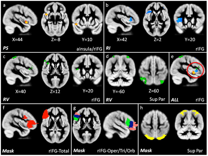
Figure 2.

Significant results (p<0.05, df 30) of grey matter morphometry correlated with the composite measures of: 2a) PS (Processing Speed; yellow/red) in the anterior insula and rIFG, 2b) RI (Response Inhibition; blue) in the rIFG, 2c) RV (Response Variability, green) in the rIFG and 2d) superior parietal lobule and 2e) an overlay of all three significant findings, highlighting the localization within rIFG (red circle) in ADHD individuals. Figure 2f–h shows the masks used to extract grey matter volume in the 2f) rIFG-Total (red), 2g) rIFG-Opercularis (green), rIFG-Triangularis (blue), rIFG-Orbitalis (pink) and 2h) the superior parietal lobule (yellow). All images are shown in radiological convention (right on left side of image).
