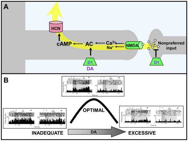
Figure 4.
DA stimulation of D1Rs weakens PFC network connections. A. Working model showing D1Rs on a different subset of spines than those containing α2A-ARs; the spines receive network inputs from neurons with dissimilar tuning characteristics (e.g. a neuron with a preferred visuospatial direction of 90° receiving an input from a neuron with a preferred response to 120°). D1R stimulation leads to cAMP generation and the opening of ion channels that shunt nearby synaptic inputs. DA and D1R are shown in purple as they decline with normal aging. B. Stimulation with the D1R agonist, SKF8129, produces an inverted-U dose-response whereby moderate, optimal doses enhance spatial tuning by reducing firing during the delay period only for the memory of nonpreferred directions. In contrast, high doses of D1R stimulation suppress firing for all directions. Adapted from [8]. F=fixation; C=cue; D=delay; R=eye movement response; PD=preferred direction of the neuron; NPD=an example of a nonpreferred direction of the neuron.

