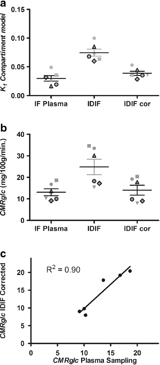
Fig. 6.
CMRglc from standard three-compartment model analyses of 18F-FDG dynamic brain imaging, using an ROI on the frontal lobe of the brain. a The K 1 fit from the compartmental model was computed using three different input functions: plasma sampling (Plasma), IDIF without correction, and IDIF corrected for carotid artery PVE (IDIF cor). b CMRglc was computed using three different input functions: plasma sampling (Plasma), IDIF without correction, and IDIF corrected for carotid artery partial volume (IDIF cor). For each subject, the diameter of the carotid artery was obtained from the bolus PET/CT coregistration images by measuring artery size on the CT image. The data from each patient are identified by the same symbol across all conditions. c Correlation between CMRglc of the frontal brain region derived from plasma sampling and from IDIF corrected for PVE

