
Figure 6.
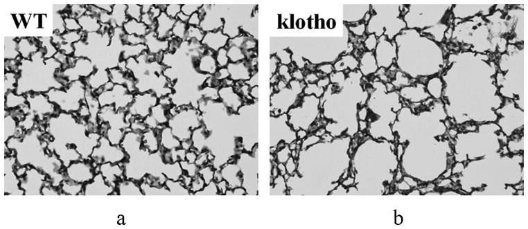
Micrographic histological images of the lung parenchyma in a WT (a) and klotho (b) mice (hematoxylin/eosine; original magnification ×400). The klotho mice exhibited larger air spaces with fewer alveolar walls, and smaller exponents in the shape and size distribution of the air spaces in the entire lobes.