
Figure 6. Electron micrographs of cardiomyocytes of papillary muscle.
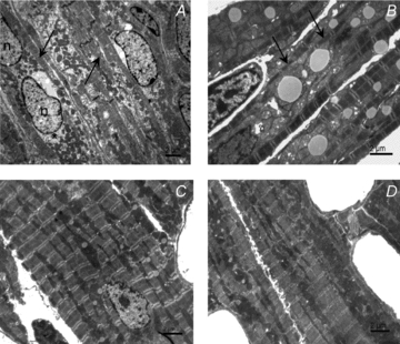
A, 3-day-old cardiomyocytes having a high content of free cytoplasm, myofibrils situated under the plasma membrane (arrows) and mitochondria clustered in the perinuclear regions (*). B, 7-day-old cardiomyocytes showing abundant mitochondrial clusters, and myofilaments of small diameters (arrows). C, 21-day-old cardiomyocytes showing regularly arranged myofilaments and mitochondria aligned in the longitudinal direction. D, 63-day-old cardiomyocytes demonstrating regular overall ultrastructure. Myofilaments and mitochondria are arranged in parallel along the longitudinal axis.