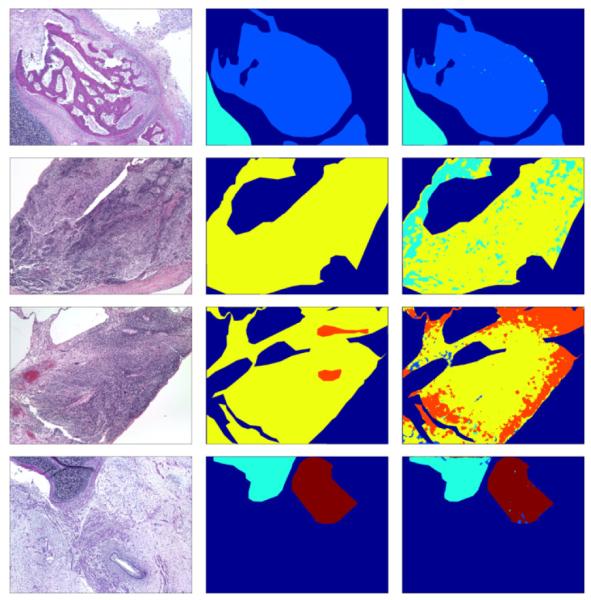
Figure 3.

Top to bottom: Example delineations of tissues for 2-, 3-, 4-, and 5-class problems respectively. Left to right: Original image, expert-labeled ground truth, our algorithm’s labeling. Color coding: B (light blue), C (cyan), I (yellow), N (orange), F (maroon), other tissues (dark blue).
