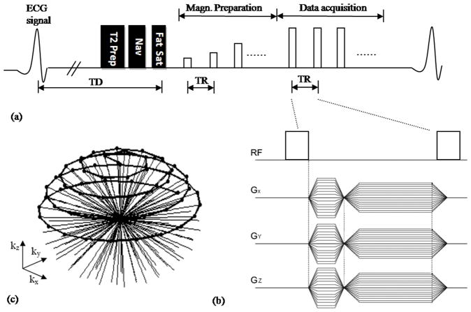
Figure 1.
(a) Schematic of the pulse sequence used for SSFP VIPR whole-heart coronary MRA. Adiabatic T2-prep, Navigator (Nav), and Spectral Presaturation Inversion Recovery (SPIR) fat saturation pulses were applied prior to imaging. Fifteen sinusoidal preparation pulses were applied prior to imaging to reduce transient signal oscillations. (b) GX, GY and GZ between RF hard pulse represent gradients along the three orthogonal axes forming the VIPR trajectory. (c) k-space coverage with the VIPR trajectory. Black dots represents signal acquired.

