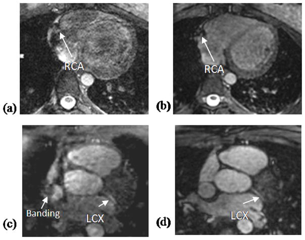
Figure 3.
Comparison between Cartesian (a, c) and VIPR (b, d) trajectory in two healthy volunteers. The Cartesian images (a, c) show severe off-resonance artifacts and poor image quality. In contrast, the VIPR images (b, d) show no obvious artifacts, have homogeneous blood pool signal, good image quality and sharp depiction of the LAD and LCX.

