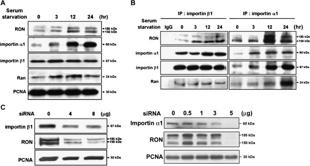
Fig. 2.
Importins/NLS-mediated nuclear import of RON. TSGH8301 cells were starved for different time periods as indicated and then nuclear extracts were collected for the following western blotting and immunoprecipitation assays with the indicated antibodies. (A) Upon serum starvation, increasing expression of RON, importin α1, importin β1 and Ran in the nucleus were detected by immunoblotting analysis. Proliferating cell nuclear antigen (PCNA) was used as a nuclear marker. (B) The association of RON with nuclear importin proteins, including importin α1, importin β1 and Ran, was determined by co-immunoprecipitation assays. Nuclear extracts were pulled down by either anti-importin β1 (left) or anti-importin α1 (right) antibodies and then probed with the indicated antibodies. (C) Inhibition of nuclear RON expression by importin α1 or β1 siRNA knockdown. TSGH8301 cells were transfected with different dosage of importin α1 or β1 siRNA for 48 h and then nuclear extracts were collected for sodium dodecyl sulfate–polyacrylamide gel electrophoresis resolution. PCNA was as a nuclear marker and loading control. A dose-dependent suppressor effect was more apparent in knock down of importin β1.

