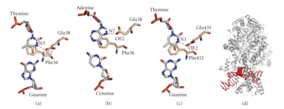
Figure 5.
Mismatch recognition mode of MutS. (a) G:T mismatch bound to E. coli MutS (PDB ID: 1E3M). (b) C:A mismatch bound to E. coli MutS (PDB ID: 1OH5). Cytosine residue is in a syn conformation. (c) G:T mismatch bound to human MutSα (PDB ID: 2O8B). (d) Side view of the E. coli MutS-mismatch complex (PDB ID: 1E3M). The mismatched duplex is sharply kinked in the complex with MutS. MutS and mismatched DNA are colored grey and red, respectively. The mismatched G and T are shown in the sphere model.

