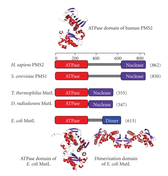
Figure 7.
(a) A schematic representation of the domain structure of MutL homologues. ATPase, nuclease, and dimer indicate the ATPase, endonuclease, and dimerization domains, respectively. The crystal structures of N-terminal ATPase domain of human PMS2 (PDB ID: 1EA6) [45], ATPase domain of E. coli MutL (PDB ID: 1B63) [46], and C-terminal dimerization domain of E. coli MutL (PDB ID: 1X9Z) [47, 48] are shown.

