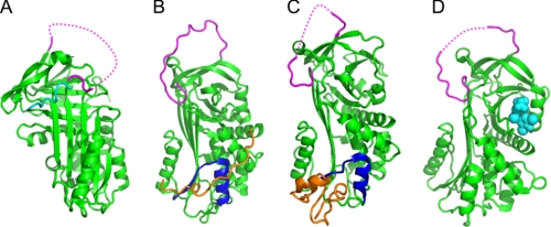
FIGURE 3.
Recent serpin structures. A, structure of native thermopin (24). The serpin is in green, with the RCL in magenta (part of the RCL is not visible in electron density; the dotted line shows connectivity). The C-terminal sequence is in addition to the serpin domain and folds across the front of β-sheet A. B, structure of native tengpin (26). The serpin is in green, with the RCL in magenta and helix E/s1A in dark blue. The N-terminal sequence (orange coil) is crucial for maintaining the native metastable state. C, structure of PAI-1 in complex with the somatomedin B domain of vitronectin (orange). The serpin is in green, with the RCL in magenta and helix E/s1A in dark blue. D, structure of native TBG in complex with thyroxine (28). The serpin is in green, the RCL is in magenta, and thyroxine is in cyan spheres.

