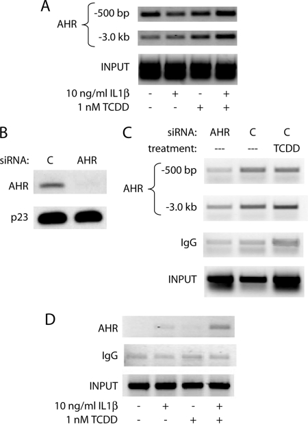
FIGURE 5.
AHR is present at the IL6 promoter in the region of the transcription start site and −3.0-kb DREs and remains over time. A, ChIP analysis of the MCF-7 IL6 promoter following 2 h of treatment with vehicle, 10 ng/ml IL1β, 1 nm TCDD, or TCDD+IL1β. AHR was immunoprecipitated, and DNA was amplified using primers for the region specified upstream from the transcription start site. B, MCF-7 cells were electroporated with control or AHR targeted siRNA oligonucleotides, plated for 24 h, and serum-starved for 18 h. Whole cell extracts were prepared, and protein levels of AHR and p23 (control) were assessed by immunoblot. C, MCF-7 cells were electroporated and serum-starved as above and then treated for 2 h with vehicle control or 10 nm TCDD. ChIP analysis of the IL6 promoter was then carried out, and DNA was amplified using primers for the region specified upstream from the transcription start site. D, ChIP analysis of the MCF-7 IL6 promoter following 6 h of treatment with vehicle, 10 ng/ml IL1β, 1 nm TCDD, or TCDD+IL1β. AHR was immunoprecipitated, and DNA in the region of −500 bp upstream from the transcription start site was amplified.

