FIGURE 4.
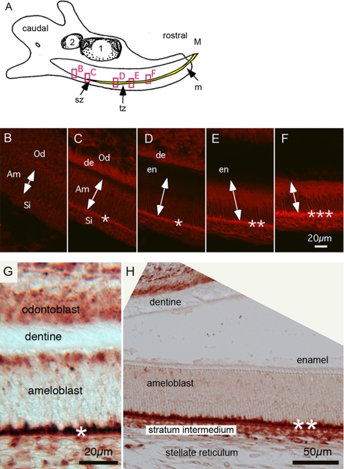
Immunofluorescence (IF) and immunoperoxidase of NBCe1 in polarized ameloblast cells. A, illustration of a sagittal view of a mandible of 3-day-old wild-type mouse. The mature (M) end and the caudal or growing end (GE) of the incisor tooth are identified. Also identified are the secretory zone (sz), transitional zone (tz; the zone between enamel matrix secretion and enamel maturation) and mature end (m) of the incisor enamel. Boxed regions from left to right are approximate regions shown in B–F. B–F, immunoflourescence of NCBe1 in a 3-day-old developing mandibular incisor. Expression of NBCe1 is seen at the various stages of amelogenesis. B, nonpolarized presecretory stage. C, early stage secretory, signal is recognizable at this point. D, secretory stage showing clear expression of NBCe1 at basolateral pole. E and F, the highest expression of NBCe1 is found in the cells at the end of the secretory stage. Double-headed arrows identify orientation and basal (lower) and apical (upper) extremes of ameloblast cells. Asterisks identify relative levels of NBCe1 expression. Am, ameloblast; Od, odontoblast; Si, Stratum intermedium; en, enamel; and de, dentin. G and H, immunoperoxidase of polarized, early secretory (G) and secretory stage (H) ameloblasts of the mandibular incisor. Note that a separation artifact between the enamel and dentin occurred during tissue preparation from immunoperoxidase staining (H). Scale bars: B–F, 20 μm; G, 20.0 μm; and H, 50.0 μm.

