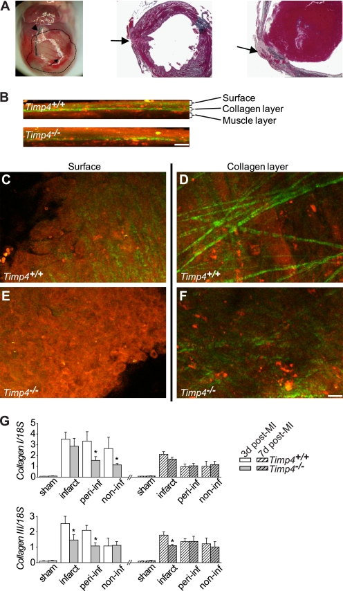
FIGURE 4.
Cardiac rupture and decreased collagen synthesis and disruption of collagen network in Timp4−/− hearts after MI. A, a representative picture of a ruptured heart following MI and trichrome-stained fixed LV tissue sections showing the rupture area (arrow) and LAD ligation (arrowhead). The infarct area is marked with a dotted line. B–F, second harmonic generation (green pseudocolor) and multiphoton fluorescence (red pseudocolor) images indicating compromised fibrillar collagen and altered cellularity in Timp4-deficient hearts post-MI. B, the depth profile extending below the heart surface. C–F, 10-μm-thick maximum intensity projections of the surface and collagen layers of Timp4+/+ and Timp4−/− heart infarct regions. Scale bar, 20 μm (B–F). G, Taqman RT-PCR analysis of RNA for collagen I and III in hearts of Timp4+/+ and Timp4−/− mice at 3 and 7 (dashed bars) days after sham operation or myocardial infarction. Values were normalized to 18 S rRNA and are expressed as mean ± S.E.; n = 4–6 for each group. *, p < 0.05 versus Timp4+/+ MI. peri-inf, peri-infarct; non-inf, noninfarct.

