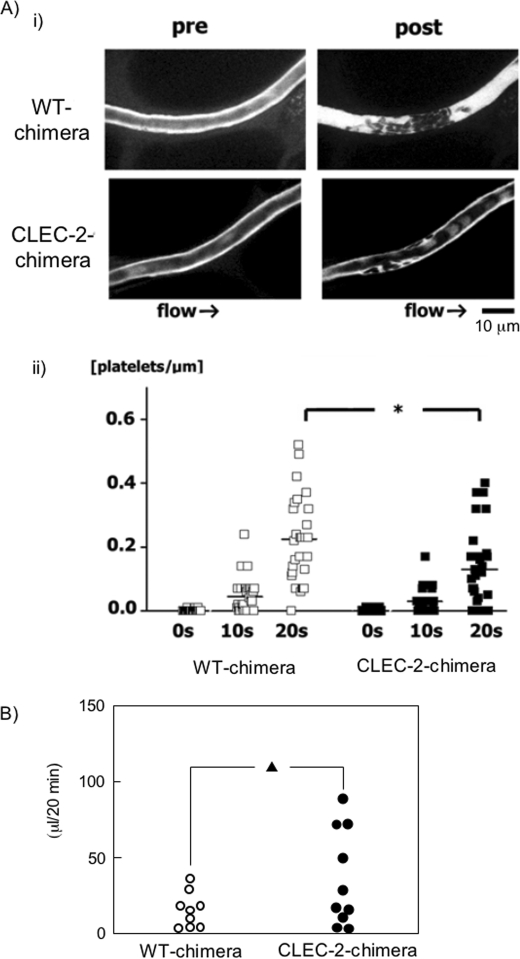
FIGURE 7.
In vivo thrombus formation in a laser-induced injury model is impaired in CLEC-2 chimeras without significant increase in tail bleeding. A, video stills of mesenteric capillaries were obtained by intravital fluorescence microscopy before (pre) and 20 s after (post) laser-induced injury (panel i). The numbers of platelets in developing thrombi after laser injury to capillaries were calculated (panel ii). The y axis represents the numbers of platelets/micrometer of obtained vessel length. Results from WT and CLEC-2 chimeras 7 weeks after transplantation (17 weeks old) are shown (n = 5 each). *, p < 0.05. B, shown is the tail bleeding in WT and CLEC-2 chimeras. Each symbol represents one individual. Results from WT and CLEC-2 chimeras 8 weeks after transplantation (17 weeks old) are shown. ▴, not significant (p = 0.08).

