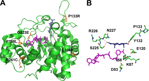
FIGURE 1.
Location of the disease-related mutants in the AKR1D1 crystal structure. A, position of the mutants on the structure and their relationship to cofactor (magenta) and steroid (blue) binding. B, a perspective view of the location of Pro133 and its relationship to binding testosterone in the unproductive binding mode.

