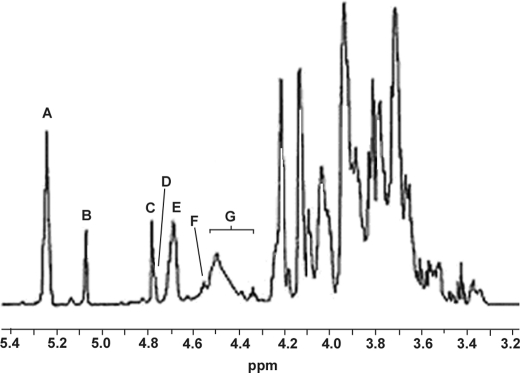
FIGURE 1.
1H NMR spectrum of Hyp-AG interferon Hyp-polysaccharide-1 at 55 °C. Signals A and B were assigned respectively to H-1 of six and two α-l-Ara residues, signal C to H-1 of two α-l-Rha residues, signal D (shoulder peak of C) to H-4 of Hyp, and signal E to the five β-d-Gal residues of the galactan backbone. Signal F was assigned to H-1 of β-d-Gal linked to Hyp and signal G to H-1 of the four side chain β-d-Gal residues and two β-d-GlcUA residues.

