FIGURE 9.
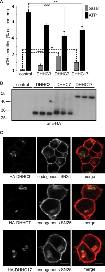
Effects of DHHC overexpression on exocytosis in PC12 cells. A, PC12 cells were co-transfected with a plasmid encoding hGH together with HA-DHHC3, DHHC7, DHHC17, or empty HA plasmid (control). Forty-eight hours post-transfection, the cells were washed and incubated in Krebs buffer in the absence (basal) and presence of 300 μm ATP. The secreted and cell-associated hGH was assayed using an enzyme-linked immunosorbent assay kit, and secretion determined as a percentage of the total cell content. Results from three independent experiments (n = 3 for each experiment) were averaged, and the means ± S.E. are presented. One-way ANOVA tests revealed that DHHC7 and DHHC17 significantly reduced ATP-stimulated secretion compared with control transfected cells. The basal secretion level for DHHC7- and DHHC17-transfected cells was significantly increased compared with control transfected cells. *, indicates a p value of < 0.05; **, denotes a p value of < 0.01; ***, is p < 0.001. B, PC12 cells transfected with the HA-DHHC constructs were lysed, separated by SDS-PAGE, and transferred to nitrocellulose for immunoblotting analysis with anti-HA. Molecular weight standards are shown on the left. C, PC12 cells transfected with HA-DHHC3, HA-DHHC7, or HA-DHHC17 were fixed and permeabilized. The cells were incubated with anti-SNAP25 (SMI81) and anti-mouse antibody conjugated to Alexa Fluor 647 to stain endogenous SNAP25 and anti-HA conjugated to Alexa Fluor 488 to stain the HA-tagged DHHC constructs. Scale bars represent 5 μm.

