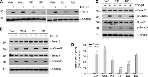
FIGURE 3.
Activation of MAPK signaling is partially responsible for PAI-1 induction by TGF-β1. HKC-8 cells were pretreated with either various chemical inhibitors or vehicle (Veh., 0.1% Me2SO) as indicated for 30 min, followed by incubation in the absence or presence of TGF-β1 (2 ng/ml) for 24 h. PD, PD98059 (MEK1 inhibitor) (10 μm); wort., wortmannin (PI3K inhibitor) (10 nm); SC, SC68376 (p38 MAPK inhibitor) (20 μm); SP, SP600125 (JNK inhibitor) (20 μm); RO, Ro31-8220 (pan-specific protein kinase C inhibitor) (10 μm). A, representative Western blots of PAI-1 expression after various treatments. B and C, Smad2/3 abundance and phosphorylation in the presence of various inhibitors as indicated. D, quantitative determination of the relative PAI-1 abundance after normalization with GAPDH. The data (relative to the controls = 1.0) are presented as the means ± S.E. of three experiments. *, p < 0.05; **, p < 0.01 versus vehicle controls.

