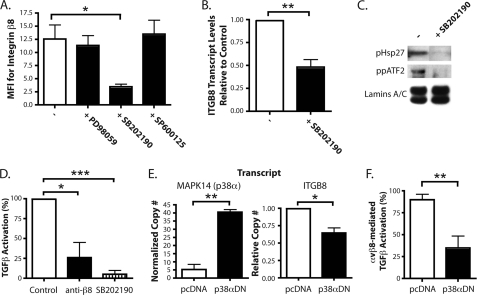
FIGURE 6.
p38 regulates ITGB8 expression and αvβ8-mediated TGF-β activation. A, flow cytometry for integrin β8 on adult lung fibroblasts treated with MAPK inhibitors, PD98059 (ERK), SB202190 (p38), and SP600125 (JNK) (± S.E.). MFI, mean fluorescence intensity. B, quantitative RT-PCR results for ITGB8 expression in adult lung fibroblasts treated with SB202190, normalized to GAPDH and β-actin and relative to control (± S.E.). C, immunoblot for phosphorylated HSP 27 and dual-phosphorylated ATF-2 from nuclear extracts from adult lung fibroblasts treated ± SB202190. Immunoblot for the nuclear localized proteins, lamins A and C, was used as a loading control. D, TGF-β activation assays of adult lung fibroblasts treated with anti-β8 blocking antibodies or SB202190 (± S.E.). E, quantitative RT-PCR results for MAPK14 (p38α) and ITGB8 in adult lung fibroblasts transfected with plasmids expressing a p38α dominant-negative isoform (p38αDN) or the empty vector control, pcDNA (± S.E.). The measured transcript is labeled above each respective graph. F, TGF-β activation assays of adult lung fibroblasts transfected with plasmids expressing a p38α dominant-negative isoform (p38αDN) or the empty vector control, pcDNA. Percentage (%) of αvβ8-mediated TGF-β activation shown (± S.E.). * = p ≤ 0.05; ** = p ≤ 0.01; *** = p ≤ 0.001.

