FIGURE 1.
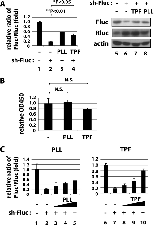
PLL and TPF suppress shRNA-mediated gene silencing. A, 293T cells were transfected with expression plasmids for firefly (Fluc) and Renilla luciferase (Rluc) together with a shRNA that targets Fluc (sh-Fluc, lanes 2–4) or a control irrelevant shRNA (sh-control, lane 1). Sh-Fluc is designed to silence Fluc, but not Rluc. In parallel, cells were treated with 3.3 μm PLL (lane 3), 3.3 μm TPF (lane 4), or were untreated (lanes 1 and 2). In the left graph, luciferase activities were quantified at 24 h post treatment, and Fluc/Rluc ratios are graphed based on the averages from three independent experiments. In the right panels, Fluc (top), Rluc (middle), and actin (bottom) as an internal control were immunoblotted. Effective silencing of Fluc by sh-Fluc was seen in lane 2 while treatment with PLL (lane 3) or TPF (lane 4) reduced the effectiveness of silencing. B, MTT assays for cell viability of control-treated or PLL- or TPF-treated cells. C, dose-dependent suppression of shRNA silencing by PLL (left) and TPF (right). 293T cells were transfected with plasmids as in A and then treated with escalating amounts of PLL (0, 0, 0.31, 1.3, and 5.0 μm in lanes 1–5) or TPF (0, 0, 1.5, 3.3, and 7.5 μm in lanes 6–10). Luciferase activities were quantified as in A.

