FIGURE 8.
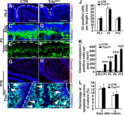
Normal proliferation and differentiation determination but increased apoptosis of Trioflox/flox; Nestin-Cre granule cells. A and B, phospho-histone-H3 (H3) immunostaining shows normal proliferation ability of mutant EGL. Dashed lines indicate the pial surface. C and D, the expression pattern for process extension can be detected by Tuj1 in the mutant inner EGL at P3. E and F, the differentiated neuron maker NeuN exists in the inner EGL in the mutant mice as the control at P3. Dashed lines indicate the pial surface. G and H, apoptosis, characterized by cleaved caspase-3, is greatly aggravated in mutant cerebella. I, most of the apoptotic cells in the mutant are NeuN-positive (white arrowheads) in the inner EGL or molecular layer. Dashed lines indicate the pial surface. J, H3-positive cells at the outer EGL per unit length of EGL. K, quantification of apoptotic cerebellar cells per area of cerebella during different developmental stages (n = 3); ***, p < 0.001. L, quantification of apoptotic granule cells in vitro by propidium iodide staining. Cerebellar neurons were from P4 mice and cultured at high density (600 neurons per mm2). The histogram indicates that the percentage of apoptotic cells in Trio mutant neurons was comparable to CTR cultured for 2 and 4 days. Scale bars in A–I, 100 μm.

