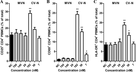
FIGURE 4.
MVN does not increase the expression of cellular activation markers. PBMCs were cultured in the presence of varying concentrations of the lectins MVN and CV-N and incubated at 37 °C. At day 3, the PBMCs were analyzed by flow cytometry for their expression of cellular activation markers with phycoerythrin-conjugated anti-CD25 (A), anti-CD69 (B), or anti-HLA-DR (C) mAbs in combination with FITC-conjugated anti-CD4 mAb. Data represent mean percentage ± S.E. for 4–16 independent experiments. *, p < 0.05; **, p < 0.0001 for comparison with untreated PBMCs (Student t test).

