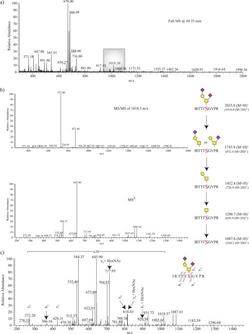
FIGURE 3.
Assignment of an O-GalNAc α-DG glycopeptide. From the full scan acquired at 49.33 min (a), a peak at 1018.3 m/z was selected for fragmentation (b). The resulting MS/MS of 1018.3 m/z yielded the neutral loss of two terminal SA residues, which was then followed by MS3 fragmentation indicating the loss of a Gal residue followed by a reducing end GalNAc. The combined glycan structure was determined to belong to a peptide with 1087.6 m/z. From examining the MS/MS spectra (not shown) and the neutral loss-triggered MS3 spectra (c), the site of post-translational modification to the serine within the peptide IRTTTSVGPR is assigned.

