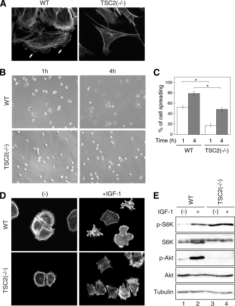
FIGURE 1.
Loss of TSC2 expression inhibits cell spreading and actin rearrangement. A, morphology of WT and TSC2(−/−) REF cells on culture plates. WT and TSC2(−/−) REF cells grown on glass coverslips were fixed and stained with Texas Red-conjugated phalloidin. Formation of lamellipodia was observed in WT cells as indicated by solid arrows. B, WT and TSC2(−/−) REF cells were serum-starved for 2 h and replated onto fibronectin-coated coverslips for 1 and 4 h. Representative phase contrast images were taken from the WT and TSC2(−/−) cells after spreading for indicated time. C, quantitative representation of summarized results obtained from three independent experiments. The percentage of cell spreading was calculated as described under “Experimental Procedures.” For statistical analysis, the data obtained from the TSC2(−/−) cells were compared with the WT cells, and the p values were determined by two-sample t tests. Data represent the mean ± S.E. (n = 3; *, p < 0.05). D, after allowing cells to spread on fibronectin-coated coverslips for 1 h, one set of the cells were treated with IGF-1 (20 ng/ml) for 5 min. The cells were subsequently fixed and stained with Texas Red-conjugated phalloidin. Images shown were obtained with a 20× objective. E, activation of PI3K signaling is attenuated in TSC2(−/−) REF cells. WT and TSC2(−/−) REF cells were serum-starved for 2 h and subsequently stimulated with IGF-1 (20 ng/ml) for 10 min. The cells lysates were prepared and analyzed for the phosphorylation status of p70S6K and AKT.

