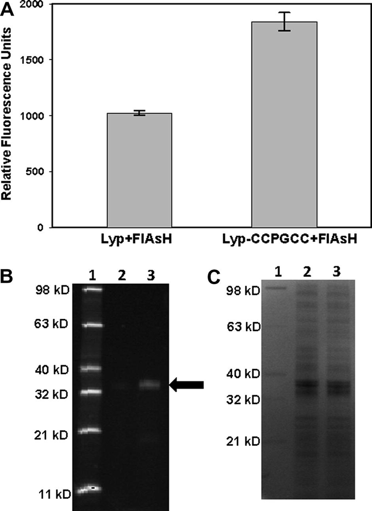
Figure 6.
Targeting Lyp-CCPGCC in E. coli cell preparations. (A) Freeze-thawed E. coli cells expressing either wild-type Lyp or Lyp-CCPGCC were incubated in the presence of FlAsH (10 µM). After 2.5 hours, the FlAsH-fluorescence values of the cell suspensions were measured. (B, C) E. coli cells expressing either Lyp or Lyp-CCPGCC were prepared and FlAsH-treated as in (A) and subsequently lysed. Cellular proteins were separated by SDS-PAGE. FlAsH-labeled proteins were detected by fluorescence (B), followed by visualization of all proteins in the same gel by Coomassie staining (C). Lane 1: Fluorescent protein standard (Invitrogen), Lane 2: Lysate from Lyp-expressing cells, Lane 3: Lysate from Lyp-CCPGCC-expressing cells. The black arrow indicates the prominent fluorescent 37-kD band that is enriched in Lyp-CCPGCC-expressing lysates.

