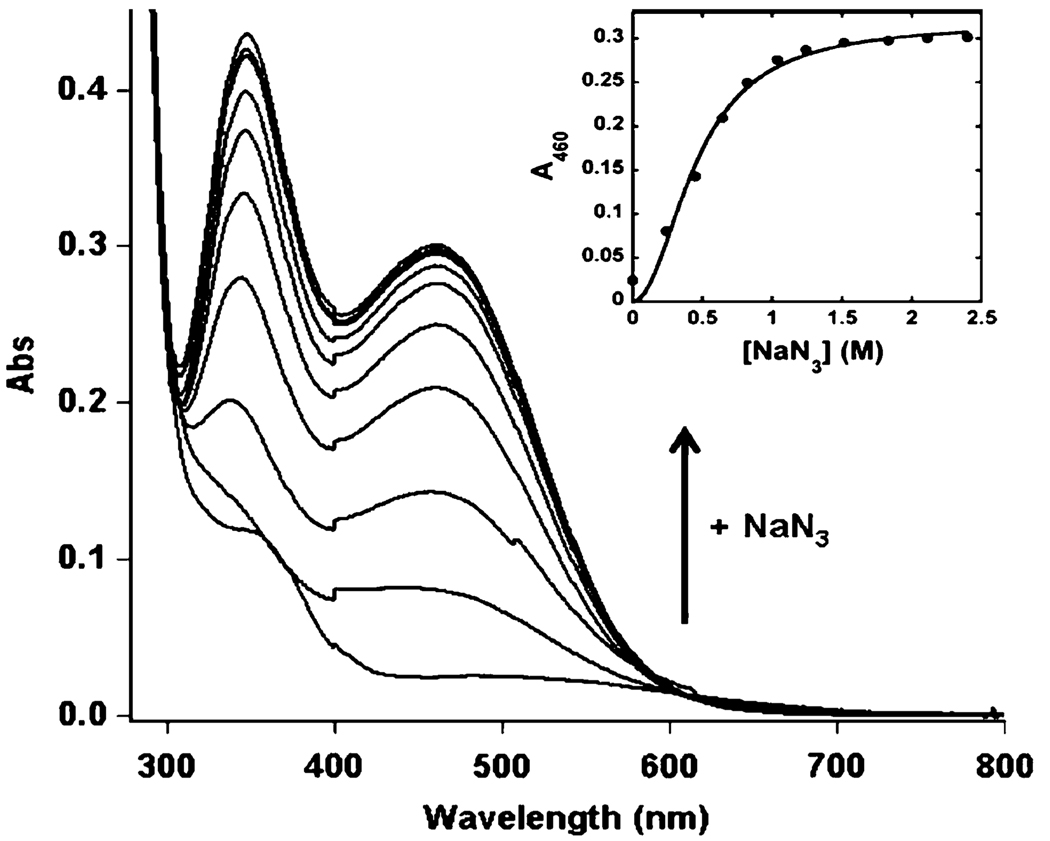
Fig. 9.
Titration of di-Fe(III)–DF3 (78 µM) with NaN3 in 50 mM HEPES, 0.1 M NaCl, pH 7 in a 1 cm path length quartz cuvette. UV– vis spectrum of di-Fe(III)–DF3 upon addition of increasing amounts of NaN3. The inset shows the increase in absorbance at 460 nm as a function of ligand concentration. The curve was obtained from a fit of the data to the Hill equation (see the text)

