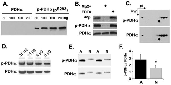
Fig. 3.
Characterization of phospho-specific antibody to serine 293 of PDHα (PDHαpS293). (A) Recombinant human PDHα and PDHα containing PDHαpS293 were probed by Western blot using the anti- PDHαpS293 serum. (B) Rat brain mitochondria were incubated with 32P-ATP in the absence or presence of 10 mM Mg2+ or 1 mM EDTA for 30 min, before analysis by Western blot. The blot was exposed to film to generate an autoradiogram for the anti-PDHαpS293 serum and then stripped and re-probed for total PDHα. (C) Rat brain mitochondria were subjected to 2D-gel electrophoresis, membrane transfer, and probed by Western blot with anti- PDHαpS293 serum. The same blot was stripped and re-probed for total PDHα. (D) Rat brain extract was probed with anti- PDHαpS293 serum, then stripped and re-probed for total PDHα. Amount of protein loaded is indicated. (E) Astrocyte and neuronal cell extracts were probed by Western blot using anti PDHαpS293 serum; the same blot was stripped and re-probed for total PDHα. (F) Immunoblots of astrocyte and neuronal cell extracts were analyzed by densitometry and expressed as the ratio of phospho-PDHα to PDHα alpha. Data presented are representative of quadruplicate measurements from three independent cultures.

